الملتحمة
| الملتحمة Conjunctiva | |
|---|---|
 النصف العلوي من مقطع سهمي من خلال مقدمة مقلة العين. (تسمية "الملتحمة" مرئية في منتصف اليسار) | |
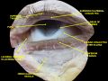
 القسم الأفقي من مقلة العين. (الملتحمة مكتوب عليها أعلى اليسار) | |
| Details | |
| جزء من | العين |
| الشريان | الشريان الدمعي, الشرايين الهدبية الامامية |
| العصب | العصب فوق البكرة |
| المُعرفات | |
| اللاتينية | tunica conjunctiva |
| MeSH | D003228 |
| TA98 | A15.2.07.047 |
| TA2 | 6836 |
| FMA | 59011 |
| المصطلحات التشريحية | |


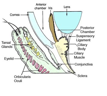
الملتحمة عبارة عن نسيج حيوي يبطن داخل الجفون ويغطي الصلبة (أبيض العين ). وهو يتألف من ظهارة حرشفية طبقية غير متقرنة مع خلايا كأسية ، وظهارة عمودية طبقية . تكون الملتحمة وعائية للغاية، مع سهولة الوصول إلى العديد من الأوعية الدقيقة للدراسات التصويرية.
التكوين
تنقسم الملتحمة عادةً إلى ثلاثة أجزاء:
| الجزء | المنطقة |
|---|---|
| الملتحمة الجسمي أو الجفن | تبطن الجفون |
| الملتحمة العينية او المقلة | يغطي مقلة العين فوق الصلبة الأمامية: هذه المنطقة من الملتحمة مرتبطة ارتباطًا وثيقًا بالصلبة الأساسية بواسطة محفظة تينون وتتحرك مع حركات مقلة العين. متوسط سماكة غشاء الملتحمة المقلة 33 ميكرون.[1] |
| الملتحمة | تشكل الوصلة بين الملتحمة المقلة وملتحمة الجفن: فهي فضفاضة ومرنة ، مما يسمح بحرية حركة الجفن ومقلة العين[2] |
إمداد الدم
الدم إلى الملتحمة المقلة يأتي في المقام الأول من الشريان العيني. يتم تدفق الدم إلى الملتحمة الجفنية (الجفن) من الشريان السباتي الخارجي. ومع ذلك ، فإن دورتي الملتحمة المقلة والملتحمة الجفنية مرتبطتان ، لذلك يتم توفير الاوعية لكل من الملتحمة المقلة والملتحمة الجفنية من كل من الشريان العيني والشريان السباتي الخارجي ، بدرجات متفاوتة..[3]
إمداد الأعصاب
ينقسم التعصيب الحسي للملتحمة إلى أربعة أجزاء:[4]
| المنطقة | العصب |
|---|---|
| علوية | |
| سفلية | العصب تحت الحجاج |
| جدارية | العصب الدمعي (بمساهمة من العصب الوجني الوجهي) |
| حول القرنية | الاعصاب الهدبية الطويلة |
التشريح الدقيق
تتكون الملتحمة من ظهارة عمودية حرشفية و طبقية حرشفية طبقية ، مع خلايا كأسية متداخلة .[5] تحتوي الطبقة الظهارية على الأوعية الدموية والأنسجة الليفية والقنوات اللمفاوية.[5] تنتج الغدد الدمعية الثانوية في الملتحمة باستمرار الجزء المائي من الدموع [5] الخلايا الإضافية الموجودة في الظهارة الملتحمة تشمل الخلايا الصباغية ، الخلايا اللمفاوية التائية و البائية.[5]
الوظيفة
تساعد الملتحمة على انزلاق العين عن طريق إنتاج المخاط والدموع ، على الرغم من حجم الدموع أقل من الغدة الدمعية.[6] كما أنه يساهم في المراقبة المناعية ويساعد على منع دخول الميكروبات إلى العين.
الأهمية السريرية
تعد اضطرابات الملتحمة والقرنية مصادر شائعة لشكاوى العين ، خاصة لأن سطح العين معرض لتأثيرات خارجية مختلفة وهو عرضة بشكل خاص للصدمات والالتهابات والتهيج الكيميائي وردود الفعل الحساسية والجفاف.
- تتأثر ديناميكا الدم للملتحمة والأوعية الدموية باعتلال الشبكية السكري (DR) ، وبالتالي يمكن أن تكون مفيدة لتشخيص DR ومراقبته,[7] والتمييز بين مراحل DR.[8]
- يرتبط داء السكري النمط الثاني بنقص الأكسجة للملتحمة,[9] وزيادة متوسط قطر الأوعية الدموية وفقدان الشعيرات الدموية.[10][11][12]
- يرتبط فقر الدم المنجلي بفقدان الأوعية الدموية ، وتغير تدفق الدم وقطر الأوعية الدموية ، والنزيف الشعري الدقيق..[13][14][15]
- يترافق فرط ضغط الدم مع زيادة في التواء الأوعية الدموية الملتحمة المقلة وفقدان الشعيرات الدموية والشرايين.[16][17]
- يرتبط انسداد الشريان السباتي بتدفق الدم الملتحمي البطيء وفقدان الشعيرات الدموية..[3]
- مع التقدم في السن ، يمكن أن تمتد الملتحمة وتخفف من صلابة الصلبة ، مما يؤدي إلى تكوين طيات الملتحمة ، وهي حالة تعرف باسم ارتخاء الملتحمة..[18][19]
- يمكن أن يتسبب داء البريميات ، وهو عدوى بالبريمية ، في حدوث اختناق الملتحمة ، الذي يتميز بوذمة الملتحمة ، والاحمرار دون الإفرازات.
الأوعية الدموية الدقيقة في الملتحمة البصلية
المورفولوجيا الوعائية
The bulbar conjunctival microvasculature contains arterioles, meta-arterioles, venules, capillaries, and communicating vessels. Vessel morphology varies greatly between subjects and even between regions of the individual eyes. In some subjects, arterioles and venules can be seen to run parallel with each other. Paired arterioles are generally smaller than corresponding venules.[20] The average bulbar conjunctival vessel has been reported to be 15.1 microns, which reflects the high number of small capillaries, which are typically <10 microns in diameter.[21]
حركة الأكسجين الدموي
The bulbar conjunctival microvasculature is in close proximity to ambient air, thus oxygen diffusion from ambient air strongly influences their blood oxygen saturation. Because of oxygen diffusion, hypoxic bulbar conjunctival vessels will rapidly reoxygenate (in under 10 seconds) when exposed to ambient air (i.e. when the eyelid is open). Closing the eyelid stops this oxygen diffusion by placing a barrier between the bulbar conjunctival microvessels and ambient air.[22]
طرق تصوير الأوعية الدموية
The bulbar conjunctival microvessels are typically imaged with a high-magnification slit lamp with green filters.[23][24][25] With such high-magnification imaging systems, it is possible to see groups of individual red blood cells flowing in vivo.[23] Fundus cameras may also be used for low-magnification wide field-of-view imaging of the bulbar conjunctival microvasculature. Modified fundus cameras have been used to measure conjunctival blood flow [26] and to measure blood oxygen saturation.[22] Fluorescein angiography has been used to study the blood flow of the bulbar conjunctiva and to differentiate the bulbar conjunctival and episcleral microcirculation.[27][28][29]
توسيع الأوعية
The bulbar conjunctival microvasculature is known to dilate in response to several stimuli and external conditions, including allergens (e.g. pollen),[30] temperature,[31] time-of-day,[31] contact-lens wear,[12] and acute mild hypoxia.[22] Bulbar conjunctival vasodilation has also been shown to correlate changes in emotional state.[32]
Type 2 diabetes is associated with an increase in average bulbar conjunctival vessel diameter and capillary loss.[10][11] Sickle-cell anemia is associated with altered average vessel diameter.[13]
انظر أيضاً
- التهاب الملتحمة (العين الوردية)
- ارتخاء الملتحمة
- جفاف العين
- شحيمة
- ظفرة
- Rougine
- نزيف تحت الملتحمة
- مرض السكري
- داء الكريات المنجلية
- مصباح شقي
معرض الصور
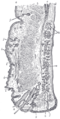
مقطع سهمي من خلال الجفن العلوي.
عضلة العين الخارجية. الأعصاب المدارية. تشريح عميق.


المصادر
- ^ Efron, Nathan; Al-Dossari, Munira; Pritchard, Nicola (2009-05-01). "In vivo confocal microscopy of the bulbar conjunctiva". Clinical & Experimental Ophthalmology. 37 (4): 335–344. doi:10.1111/j.1442-9071.2009.02065.x. ISSN 1442-9071. PMID 19594558.
- ^ Eye, human Encyclopædia Britannica
- ^ أ ب PAVLOU AT; WOLFF HG (1959-07-01). "THe bulbar conjunctival vessels in occlusion of the internal carotid artery". Archives of Internal Medicine. 104 (1): 53–60. doi:10.1001/archinte.1959.00270070055007. ISSN 0888-2479.
- ^ "Table 1: Summary of sensory nerve supply". Archived from the original on February 14, 2013. Retrieved July 31, 2016.
- ^ أ ب ت ث Goldman, Lee. Goldman's Cecil Medicine (24th ed.). Philadelphia: Elsevier Saunders. p. 2426. ISBN 978-1437727883.
- ^ London Place Eye Center (2003). Conjunctivitis Archived 2004-08-08 at the Wayback Machine. Retrieved July 25, 2004.
- ^ Khansari, Maziyar M.; Wanek, Justin; Tan, Michael; Joslin, Charlotte E.; Kresovich, Jacob K.; Camardo, Nicole; Blair, Norman P.; Shahidi, Mahnaz (7 April 2017). "Assessment of Conjunctival Microvascular Hemodynamics in Stages of Diabetic Microvasculopathy". Scientific Reports. 7: 45916. Bibcode:2017NatSR...745916K. doi:10.1038/srep45916. PMC 5384077. PMID 28387229.
- ^ Khansari, Maziyar M.; O’Neill, William; Penn, Richard; Chau, Felix; Blair, Norman P.; Shahidi, Mahnaz (1 July 2016). "Automated fine structure image analysis method for discrimination of diabetic retinopathy stage using conjunctival microvasculature images". Biomedical Optics Express (in الإنجليزية). 7 (7): 2597–2606. doi:10.1364/BOE.7.002597. ISSN 2156-7085. PMC 4948616. PMID 27446692.
- ^ Isenberg, S. J.; McRee, W. E.; Jedrzynski, M. S. (1986-10-01). "Conjunctival hypoxia in diabetes mellitus". Investigative Ophthalmology & Visual Science. 27 (10): 1512–1515. ISSN 0146-0404. PMID 3759367.
- ^ أ ب Fenton, B. M.; Zweifach, B. W.; Worthen, D. M. (1979-09-01). "Quantitative morphometry of conjunctival microcirculation in diabetes mellitus". Microvascular Research. 18 (2): 153–166. doi:10.1016/0026-2862(79)90025-6. ISSN 0026-2862. PMID 491983.
- ^ أ ب Ditzel, Jørn (1967-01-12). "The in Vivo Reactions of the Small Blood Vessels to Diabetes Mellitus". Acta Medica Scandinavica (in الإنجليزية). 182 (S476): 123–134. doi:10.1111/j.0954-6820.1967.tb12691.x. ISSN 0954-6820.
- ^ أ ب Cheung, A. T.; Ramanujam, S.; Greer, D. A.; Kumagai, L. F.; Aoki, T. T. (2001-10-01). "Microvascular abnormalities in the bulbar conjunctiva of patients with type 2 diabetes mellitus". Endocrine Practice. 7 (5): 358–363. doi:10.4158/EP.7.5.358. ISSN 1530-891X. PMID 11585371.
- ^ أ ب Fink, A I (1968-01-01). "Vascular changes in the bulbar conjunctiva associated with sickle-cell disease: some observations on fine structure". Transactions of the American Ophthalmological Society. 66: 788–826. ISSN 0065-9533. PMC 1310317. PMID 5720854.
- ^ Isenberg, S. J.; McRee, W. E.; Jedrzynski, M. S.; Gange, S. N.; Gange, S. L. (1987-01-01). "Effects of sickle cell anemia on conjunctival oxygen tension and temperature". Archives of Internal Medicine. 147 (1): 67–69. doi:10.1001/archinte.147.1.67. ISSN 0003-9926. PMID 3800533.
- ^ Wanek, Justin; Gaynes, Bruce; Lim, Jennifer I.; Molokie, Robert; Shahidi, Mahnaz (2013-08-01). "Human bulbar conjunctival hemodynamics in hemoglobin SS and SC disease". American Journal of Hematology (in الإنجليزية). 88 (8): 661–664. doi:10.1002/ajh.23475. ISSN 1096-8652. PMC 4040222. PMID 23657867.
- ^ Harper, Robert N.; Moore, Michael A.; Marr, Melissa C.; Watts, L. Earl; Hutchins, Phillip M. (1978-11-01). "Arteriolar rarefaction in the conjunctiva of human essential hypertensives". Microvascular Research. 16 (3): 369–372. doi:10.1016/0026-2862(78)90070-5.
- ^ Lee, R. E. (1955-08-01). "Anatomical and physiological aspects of the capillary bed in the bulbar conjunctiva of man in health and disease". Angiology. 6 (4): 369–382. doi:10.1177/000331975500600408. ISSN 0003-3197. PMID 13275744.
- ^ "Conjunctivochalasis - Medical Definition". Medilexicon.com. Archived from the original on 2016-03-03. Retrieved 2012-11-13.
- ^ WL Hughes Conjunctivochalasis. American Journal of Ophthalmology 1942
- ^ Meighan SS (September 1956). "Blood vessels of the bulbar conjunctiva in man". The British Journal of Ophthalmology. 40 (9): 513–26. doi:10.1136/bjo.40.9.513. PMC 1324675. PMID 13364178.
- ^ Shahidi M, Wanek J, Gaynes B, Wu T (March 2010). "Quantitative assessment of conjunctival microvascular circulation of the human eye". Microvascular Research. 79 (2): 109–13. doi:10.1016/j.mvr.2009.12.003. PMC 3253734. PMID 20053367.
- ^ أ ب ت MacKenzie LE, Choudhary TR, McNaught AI, Harvey AR (August 2016). "In vivo oximetry of human bulbar conjunctival and episcleral microvasculature using snapshot multispectral imaging" (PDF). Experimental Eye Research. 149: 48–58. doi:10.1016/j.exer.2016.06.008. PMID 27317046. S2CID 25038785.
- ^ أ ب van Zijderveld R, Ince C, Schlingemann RO (May 2014). "Orthogonal polarization spectral imaging of conjunctival microcirculation". Graefe's Archive for Clinical and Experimental Ophthalmology. 252 (5): 773–9. doi:10.1007/s00417-014-2603-9. PMID 24627137. S2CID 1595902.
- ^ Khansari MM, O'Neill W, Penn R, Chau F, Blair NP, Shahidi M (July 2016). "Automated fine structure image analysis method for discrimination of diabetic retinopathy stage using conjunctival microvasculature images". Biomedical Optics Express. 7 (7): 2597–606. doi:10.1364/BOE.7.002597. PMC 4948616. PMID 27446692.
- ^ Khansari MM, Wanek J, Felder AE, Camardo N, Shahidi M (February 2016). "Automated Assessment of Hemodynamics in the Conjunctival Microvasculature Network". IEEE Transactions on Medical Imaging. 35 (2): 605–11. doi:10.1109/TMI.2015.2486619. PMC 4821773. PMID 26452274.
- ^ Jiang H, Ye Y, DeBuc DC, Lam BL, Rundek T, Tao A, et al. (January 2013). "Human conjunctival microvasculature assessed with a retinal function imager (RFI)". Microvascular Research. 85: 134–7. doi:10.1016/j.mvr.2012.10.003. PMC 3534915. PMID 23084966.
- ^ Meyer PA (1988-01-01). "Patterns of blood flow in episcleral vessels studied by low-dose fluorescein videoangiography". Eye. 2 ( Pt 5) (5): 533–46. doi:10.1038/eye.1988.104. PMID 3256492.
- ^ Ormerod LD, Fariza E, Webb RH (1995-01-01). "Dynamics of external ocular blood flow studied by scanning angiographic microscopy". Eye. 9 ( Pt 5) (5): 605–14. doi:10.1038/eye.1995.148. PMID 8543081.
- ^ Meyer PA, Watson PG (January 1987). "Low dose fluorescein angiography of the conjunctiva and episclera". The British Journal of Ophthalmology. 71 (1): 2–10. doi:10.1136/bjo.71.1.2. PMC 1041073. PMID 3814565.
- ^ Horak F, Berger U, Menapace R, Schuster N (September 1996). "Quantification of conjunctival vascular reaction by digital imaging". The Journal of Allergy and Clinical Immunology. 98 (3): 495–500. doi:10.1016/S0091-6749(96)70081-7. PMID 8828525.
- ^ أ ب Duench S, Simpson T, Jones LW, Flanagan JG, Fonn D (June 2007). "Assessment of variation in bulbar conjunctival redness, temperature, and blood flow". Optometry and Vision Science. 84 (6): 511–6. doi:10.1097/OPX.0b013e318073c304. PMID 17568321. S2CID 943038.
- ^ Provine RR, Nave-Blodgett J, Cabrera MO (2013-11-01). "The Emotional Eye: Red Sclera as a Uniquely Human Cue of Emotion". Ethology (in الإنجليزية). 119 (11): 993–998. Bibcode:2013Ethol.119..993P. doi:10.1111/eth.12144. ISSN 1439-0310.
وصلات خارجية
- Medicinenet.com (1999). Conjunctiva. Retrieved July 25, 2004.
- MedEd at Loyola medicine/pulmonar/images/anatomy/eyeli.jpg