مهماز الصلبة Scleral spur
| Scleral spur | |
|---|---|
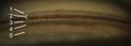
 Enlarged general view of the iridial angle (Scleral spur appear at upper right) | |
| Details | |
| الجهاز | Visual system |
| المُعرفات | |
| اللاتينية | Calcar sclerae |
| المصطلحات التشريحية | |
مهماز الصلبة scleral spur في الجهاز البصري هو نتوء في الصلبة (بياض العين) في غرفة العين الأمامية. المهماز هو بنية حلقية تتكون من كولاجين في عين الإنسان.
وهو أصل الألياف الطولية والدائرية (which swerve acutely from the spur to run circumferentially, as a sphincter near the periphery of the lens)[1] of the ciliary muscle، وترتبط من الخلف بالشبكة الحويجزية.
الدور في علاج الزرق
Open-angle glaucoma (OAG) and closed-angle glaucoma (CAG) may be treated by muscarinic receptor agonists (e.g., pilocarpine), which cause rapid miosis and contraction of the ciliary muscles, this pulls the scleral spur and results in the trabecular meshwork being stretched and separated.
This opens the fluid pathways, and facilitates drainage of the aqueous humour, into the canal of Schlemm and ultimately results in decreasing of the intraocular pressure.[2]
صور إضافية

تنظير الزاوية لزاوية الغرفة الأمامية
تنظير الزاوية لزاوية الغرفة الأمامية. البنى المسماة: 1. خط شوالبه، 2. الشبكة الحويجزية (TM), 3. مهماز الصلبة، 4. الجسم الهدبي، 5. القزحية

مقطع عرضي لزاوية الغرفة الأمامية، مصوّر باستخدام SD-OCT.

.png&filetimestamp=20200824173014&)
انظر أيضاً
المراجع
- ^ Gray's Anatomy: The Anatomical Basis of Clinical Practice (41st ed.). Elsevier. 2015. p. 693. ISBN 978-0-7020-5230-9.
- ^ Neal, M. J. (2004). "Medical Pharmacology at a Glance" (6th edition). Wiley-Blackwell. p. 27. ISBN 978-1-4051-5044-6